


Faculty
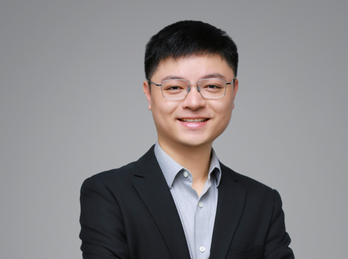
Self-introduction:
Xuesong Li is an Assistant Professor and Ph.D. supervisor in the School of Life Sciences at Southern University of Science and Technology (SUSTech). He received his B.Eng. degree from the Department of Optical Engineering at Zhejiang University in 2013 and his Ph.D. degree in Electronic and Computer Engineering from the Hong Kong University of Science and Technology (HKUST) in 2018. From 2018 to 2022, he conducted his postdoctoral research under the supervision of Dr. Hari Shroff in the High Resolution Optical and Imaging (HROI) Section with the National Institute of Biomedical Imaging and Bioengineering (NIBIB) at the National Institutes of Health, in Bethesda, Maryland, USA. From 2022 to 2026, he was a Research Specialist at Janelia Research Campus of the Howard Hughes Medical Institute (HHMI), in Ashburn, Virginia, USA. In August 2026, he joined the School of Life Sciences at SUSTech as a full-time faculty member.
His research focuses on the development of advanced optical microscopy techniques, with a particular emphasis on achieving high-spatiotemporal-resolution imaging of living biological specimens. His research interests include super-resolution fluorescence microscopy, deep-learning-enabled computational imaging, and multimodal nonlinear microscopy. He has published more than 20 peer-reviewed papers in leading international journals, including Nature Biotechnology, Nature Methods, Nature Communications, Journal of the American Chemical Society (JACS), and Proceedings of the National Academy of Sciences (PNAS). He also serves as a reviewer for leading scientific journals, including Nature Methods and Nature Communications.
Research Interests:
1. Multi-view High-Spatiotemporal-Resolution Microscopy
2. AI-Enabled Computational Microscopy
3. 3D Structured Illumination Super-Resolution Microscopy
4. Development of Light-Sheet, Confocal, and Photon-Reassignment Microscopy
Professional Experience:
◆ 2026.08 - present: School of Life Sciences, Southern University of Science and Technology, Assistant Professor.
◆ 2022.09 - 2026.07,Janelia Research Campus of the Howard Hughes Medical Institute (HHMI), Ashburn, Virginia, USA, Research Specialist
◆ 2018.10 - 2022.09: National Institute of Biomedical Imaging and Bioengineering (NIBIB) at the National Institutes of Health (NIH), Bethesda, Maryland, USA, Postdoctoral Visiting Fellow
Educational Background:
◆ 2013.09 - 2018.07: Ph.D., Electronic and Computer Engineering, The Hong Kong University of Science and Technology
◆ 2009.09 - 2013.06: B.Eng., Optical Engineering, Zhejiang University
Selected Publication:
1. Li, X.*, Wu, Y.*, Su, Y., Rey-Suarez, I.A., Matthaeus, C., Updegrove, T.B., Wei, Z., Zhang, L., Sasaki, H., Li, Y., Guo, M., Giannini, J.P., Vishwasrao, H.D., Chen, J., Lee, S-J. J., Shao, L., Liu, H., Ramamurthi, K.S., Taraska, J.W., Upadhyaya, A., La Riviere, P., Shroff, H. Three-dimensional structured illumination microscopy with enhanced axial resolution. Nature Biotechnology, 41(9), 1307-1319 (2023).
2. Li, X., Jiang, M., Lam, J. W., Tang, B. Z., & Qu, J. Y., Mitochondrial imaging with combined fluorescence and stimulated Raman scattering microscopy using a probe of the aggregation-induced emission characteristic. Journal of the American Chemical Society, 139(47), 17022-17030 (2017).
3. Li, X., Li, Y., Jiang, M., Wu, W., He, S., Chen, C., Qin, Z., Tang, B.Z., Mak, H.Y. and Qu, J.Y., Quantitative imaging of lipid synthesis and lipolysis dynamics in Caenorhabditis elegans by stimulated Raman scattering microscopy. Analytical chemistry, 91(3), 2279-2287 (2019)
4. Li, Y., Huang, S., Chan, L.S., Li, X., To, N.S., Zhao, X., Jung, K., Yang, X., Tse, Y.C., Xu, N., Xia, Y., Cheung T.H.; Qu, J.Y. and Mak, H.Y. A conserved ER protein prevents lipotoxicity by stimulating the key enzyme in glycerolipid synthesis. Nature Communications, 17, 6165 (2026)
5. Chareyre, S., Li, X., Anjuwon-Foster, B.R., Updegrove, T.B., Clifford, S., Brogan, A.P., Su, Y., Zhang, L., Chen, J., Shroff, H. and Ramamurthi, K.S. Cell division machinery drives cell-specific gene activation during differentiation in Bacillus subtilis. Proceedings of the National Academy of Sciences, 121(13), e2400584121 (2024).
6. Li, Y., Su, Y., Guo, M., Han, X., Liu, J., Vishwasrao, H., Li, X., Christensen, R., Sengupta, T., Moyle, M., Chen, J., Usdin, T.B., Colón-Ramos, D., Liu, H., Wu, Y., Shroff, H. Incorporating the image formation process into deep learning improves network performance. Nature Methods, 19(11), 1427-1437 (2022).
7. Wu, W., He, S., Wu, J., Chen, C., Li, X., Liu, K. and Qu, J.Y. Long-term in vivo imaging of mouse spinal cord through an optically cleared intervertebral window. Nature communications, 13(1), 1959 (2022).
8. Zeng, L., Li, X., Preusch, C.B., He, G.J., Xu, N., Cheung, T.H., Qu, J. and Mak, H.Y. Nuclear receptors NHR-49 and NHR-79 promote peroxisome proliferation to compensate for aldehyde dehydrogenase deficiency in C. elegans. PLoS Genetics, 17(7), e1009635 (2021).
9. Chen, J., Sasaki, H., Lai, H., Su, Y., Liu, J., Wu, Y., Zhovmer, A., Combs, C.A., Rey-Suarez, I., Chang, H., Huang, C.C., Li, X., Guo, M., Nizambad, S., Upadhyaya, A., S-J. J. Lee, Lucas, L.A.G., Shroff, H. Three-dimensional residual channel attention networks denoise and sharpen fluorescence microscopy image volumes. Nature Methods, 18(6), 678-687 (2021).